Extra-follicular Variant of Adenomatoid Odontogenic Tumor: A Diagnostic Enigma
DOI:
https://doi.org/10.3126/jbpkihs.v5i2.43747Keywords:
Adenomatoid odontogenic tumor, Cone-beam computed tomography, Extra-follicular, SwellingAbstract
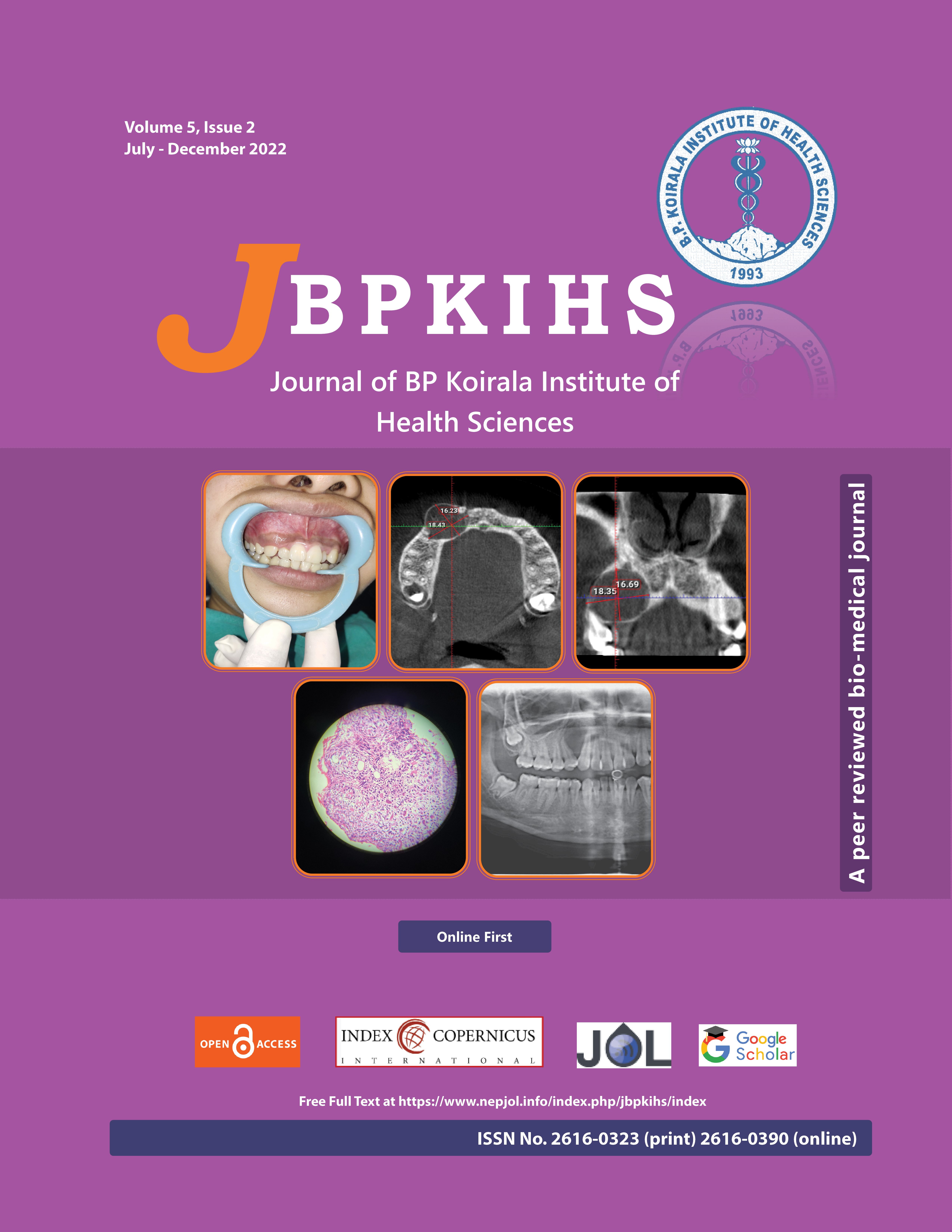
Adenomatoid odontogenic tumor (AOT) is an uncommon, slow-growing, noninvasive odontogenic tumor mostly in the anterior maxilla with three well-recognized clinico-pathological variants: follicular, extra-follicular, and peripheral. Extra-follicular variant presents as a well-defined, unilocular, radiolucency in between, above, or superimposed on the roots of an erupted tooth. A 19 years female reported with the chief complaint of a loose tooth in the right front region of the upper jaw for 6 months, associated with firm swelling without pain or discharge. On orthopantomogram and cone-beam computed tomography, the lesion appeared as a single, localized, well-defined, roughly oval unilocular radiolucency with flecks of radiopacity integrating radicular and cervical third of 13. Complete surgical enucleation followed by histopathological examination revealed the lesion as AOT.
The extra-follicular AOT can cause diagnostic dilemmas and is often misdiagnosed as an odontogenic cyst.
Downloads
Downloads
Published
How to Cite
Issue
Section
License
Copyright (c) 2022 Journal of BP Koirala Institute of Health Sciences

This work is licensed under a Creative Commons Attribution-NonCommercial-NoDerivatives 4.0 International License.
This license enables reusers to copy and distribute the material in any medium or format in unadapted form only, for noncommercial purposes only, and only so long as attribution is given to the creator.


