An overlap of Coats and Eales diseases or a Coats variant?
DOI:
https://doi.org/10.3126/nepjoph.v6i1.10782Keywords:
Coats disease, Eales diseaseAbstract
Objective: To report an association between Coats and Eales diseases, an uncommon presentation.
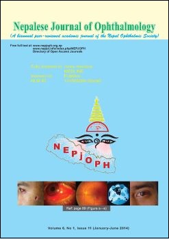
Case: A 21-year-old male presented with gradual visual impairment of two years duration in his left eye. The slit-lamp examination of the affected eye revealed +2 vitreous cells. The other findings were peri-papillary fluid accumulation and extensive macular lipid exudate deposition. Small white vessels were coursing over the macula. The major veins were dilated and tortuous and massive sheathing of both arteries and veins was forming a common sheath. In the mid-periphery and periphery of the retina, discrete hard exudates, tiny superficial retinal hemorrhages and massive vascular sheathing were present. In the inferotemporal region, two intra-retinal macrocysts were located distal to the retinal vasculature. Fluorescein angiography (FA) of the left eye highlighted numerous aneurysmal dilatations throughout the posterior pole. Fluorescein angiography also showed para-foveal telangiectasia and tiny telangiectatic vessels on the optic disk that led to late staining of the macula and optic disk. Hyperfluorescent patches of deep choroiditis were present in the early phases. There was segmental but no diffuse staining of the retinal veins which showed massive sheathing on fundoscopy. In the periphery, segmental venous staining and choroidal leakage to a lesser extent were observed. In the infero-temporal quadrant, a clear-cut zone of non-perfusion and vascular abnormalities (micro-macro aneurysms, veno-venous shunts, venous beading) at the junction between the perfused and non-perfused zones were present. The findings were reminiscent of both Coats and Eales diseases.
Conclusion: Though known as two distinct entities, both retinal pathologies may present in a single form.
DOI: http://dx.doi.org/10.3126/nepjoph.v6i1.10782
Nepal J Ophthalmol 2014; 6 (2): 109-112
Downloads
Downloads
Published
How to Cite
Issue
Section
License
This license enables reusers to copy and distribute the material in any medium or format in unadapted form only, for noncommercial purposes only, and only so long as attribution is given to the creator.


